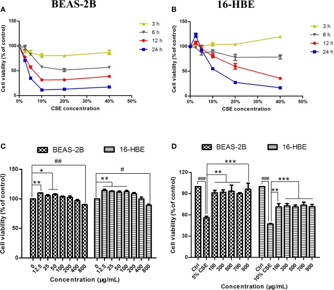
Figure 2.
Effects of CSE and JPYF II on bronchial epithelial cell viability. (A, B) The MTT assay showed the effects of different concentrations of CSE at different time on BEAS-2B and 16-HBE cell viability. (C) The MTT assay showed the effects of JPYF II on BEAS-2B and 16-HBE cell viability. Results are presented by 3 independent experiments (n = 3). Values are presented as means ± SD. *P < 0.05, **P < 0.01, #P < 0.05 and ##P < 0.01 compared with 0 μg/ml JPYF II group. (D) The MTT assay showed the effects of JPYF II on CSE-stimulated BEAS-2B and 16-HBE cell viability. Results are presented by three independent experiments (n = 3). Values are presented as means ± SD. ###P < 0.001 compared with control group; **P < 0.01 and ***P < 0.001 compared with CSE group.

