
Fig.1. AIP1A was downregulated and AIP1B was upregulated in diseased aortae.
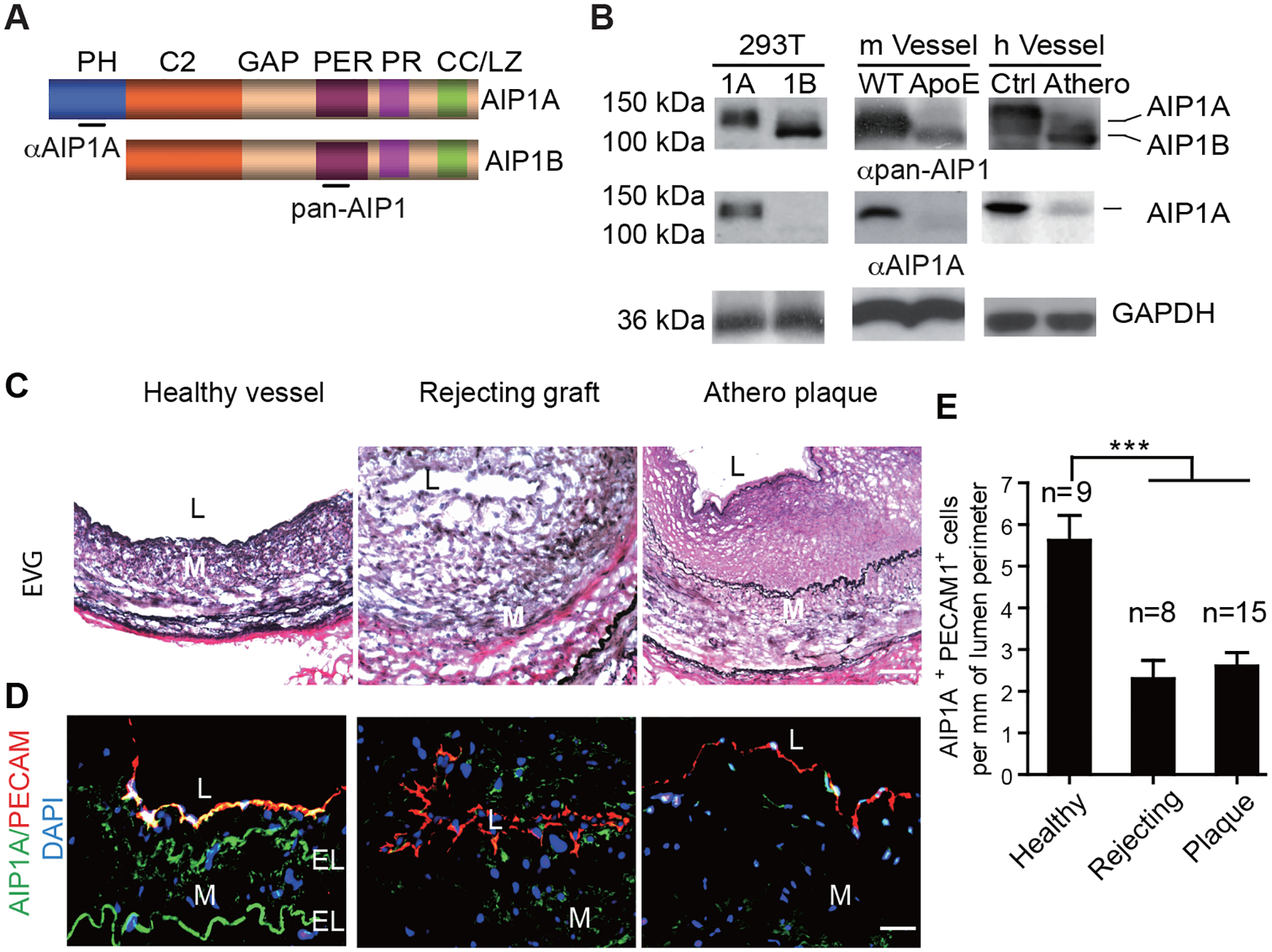
A. A schematic diagram of AIP1 structural domains. PH, PH domain; C2, PKC conserved domain; GAP, GTPase-activating protein domain; PER, period-like domain; PR: proline-rich region; CC/LZ, coiled coil/leucine-zipper domain. Antibodies that recognize the PH domain (αAIP1A) or the PER domain (αpan-AIP1) are indicated. B. Expression of AIP1A and AIP1B in the aorta. 293T: Verification of antibody specificity using lysates from 293T cells that overexpressed AIP1A or AIP1B. m Vessel: Aortae from WT and ApoE−/− mice after 20 weeks on a high-fat diet. h Vessel: human coronary artery specimens from individuals with atheroma or no disease were collected. Cell and tissues lysates were analyzed by western blotting with anti-AIP1A and anti-pan-AIP1 antibodies. n=3. C-E. Human coronary artery specimens with no disease, with graft arteriosclerosis (GA) from chronically rejecting heart allografts, or with atherosclerotic plaques were collected. EVG staining (C) and immunostaining for AIP1A expression with antibodies against AIP1A (green) and the endothelial cell marker, PECAM-1 (red). Nuclei are labelled with DAPI. Vessel lumen (L), media (M) and elastic lamina (EL) are indicated. Representative images are presented in (C) and (D) with quantification in (E). All data are presented as the mean ± SEM from independent clinical specimens of varying number per group as indicated, *** P<0.001, one-way ANOVA followed by Bonferroni’s post-hoc test. Scale bar: 50 μm (C,D).