
Fig.5. The AIP1B transgene increased carotid ligation-induced oxidative stress and inflammation.
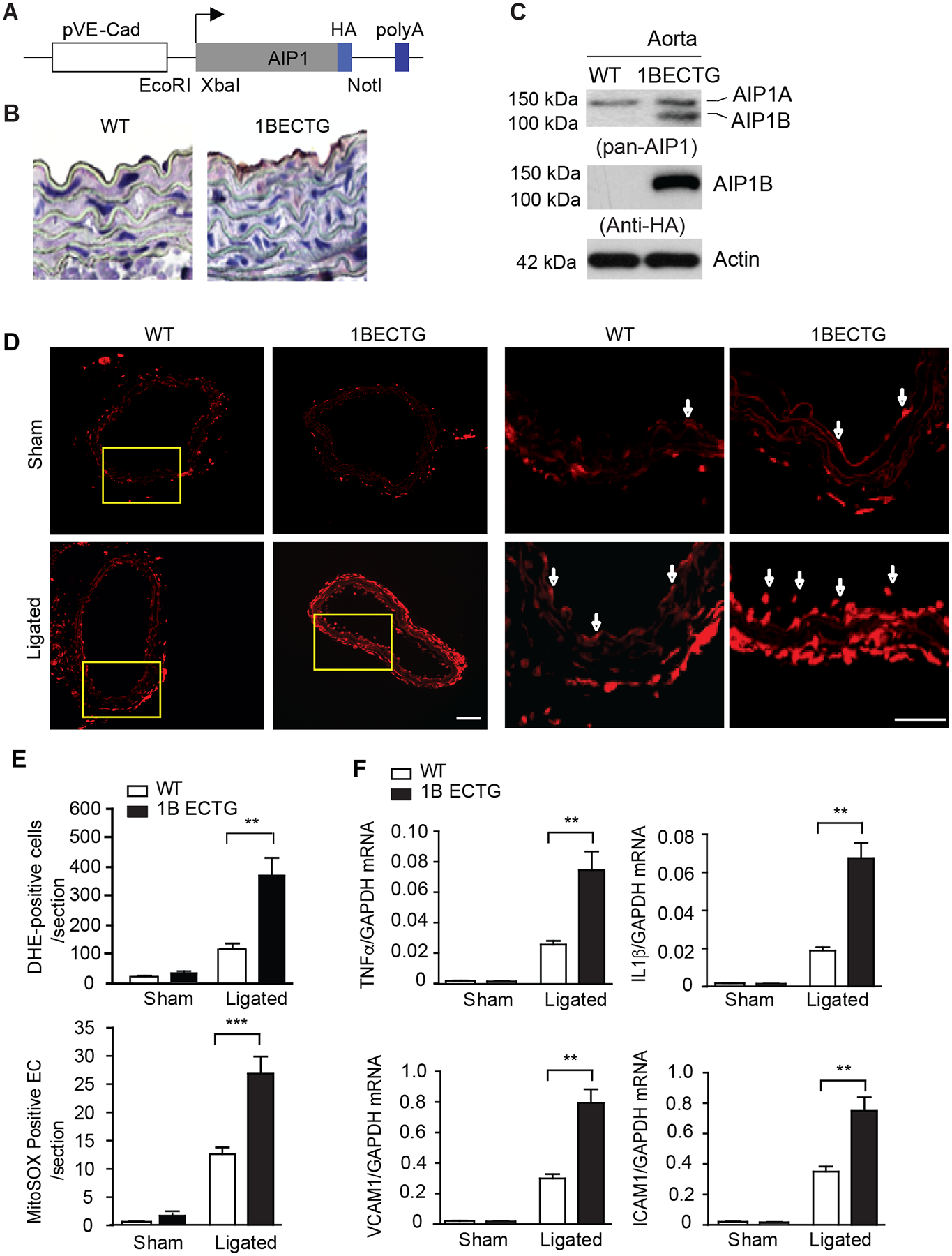
A-C. Characterization of AIP1B-ECTG mice. (A) A schematic of the transgenic vector for HA-tagged human AIP1B under the EC-specific VE-cadherin gene promoter. Restriction sites for cloning XbaI and NotI are indicated. (B) AIP1B expression in the aorta. Representative mouse aortae from WT and AIP1B-ECTG mice were collected as frozen sections, and the transgene was detected by immunohistochemistry with an anti-HA antibody. (C) Aortic AIP1 expression was detected by western blotting with anti-pan-AIP1 and anti-HA antibodies. n=2. D-F. ROS production and inflammation in AIP1B-ECTG mice. Carotid arteries and tissue from WT and AIP1B-ECTG mice with complete ligation near the carotid bifurcation on the left common carotid artery were harvested at day 3 post-ligation. Oxidative stress in the common carotid arteries was determined by in situ detection of superoxide with MitoSOX fluorescence. Representative images are shown in (D) with high magnification images of the boxed area on the right. Quantification of MitoSOX positive cells/sections are presented in (E). n=4. (F) Gene expression of inflammatory molecules was determined by RT-PCR with normalization to GAPDH. Relative expression levels are presented. n=6. All data are presented as the mean ± SEM. ** P<0.01, *** P<0.001, unpaired, two tailed t-test. Scale bar: 200 μm (D).