
Fig. 5. Immunogold labeling of DNs with ATG9A, LAC3 and RTN3 antibodies.
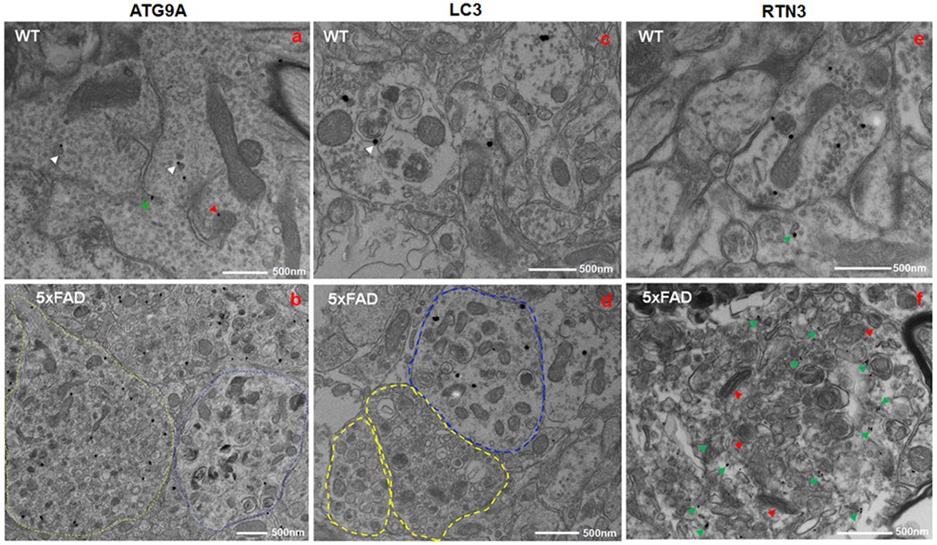
Fixed brain samples of a 6-month-old wild type (WT) (a, c and e) and a 5xFAD (b, d and f) mouse were subjected to immune-EM examination. ATG9A antibody labeled several vesicles (white arrowhead), ER tubules (green arrowhead), and mitochondrial edge (red arrowhead) in WT mouse brain (a). In 5xFAD mouse brain, ATG9A antibody stained a multi-vesicle body with small vesicles (type I DN- indicated by yellow circle), while large double membrane vesicles (type II DN- encircled in blue) were rarely detected by ATG9A antibody (b). A monoclonal antibody specific to LC3B stained isolated vesicles, styptic vesicles, and auto-lysosome-like structures (showed by white arrowhead) in a neurite in WT mouse brain (c). In 5xFAD mouse brain, LC3 localization was limited to type II DNs (d). In WT mice brain, RTN3 antibody labeled the mitochondrial edge, synaptic vesicles, and ER tubules (shown by green arrowhead) in a neurite (e). In 5xFAD mouse brain, RTN3 antibody stained tubular ER in a mitochondria (indicated by red arrowhead) – ER clustered inclusion (f).