
Fig 6. Link between ER tubules and multi-vesicles in axons of 5xFAD mouse brain.
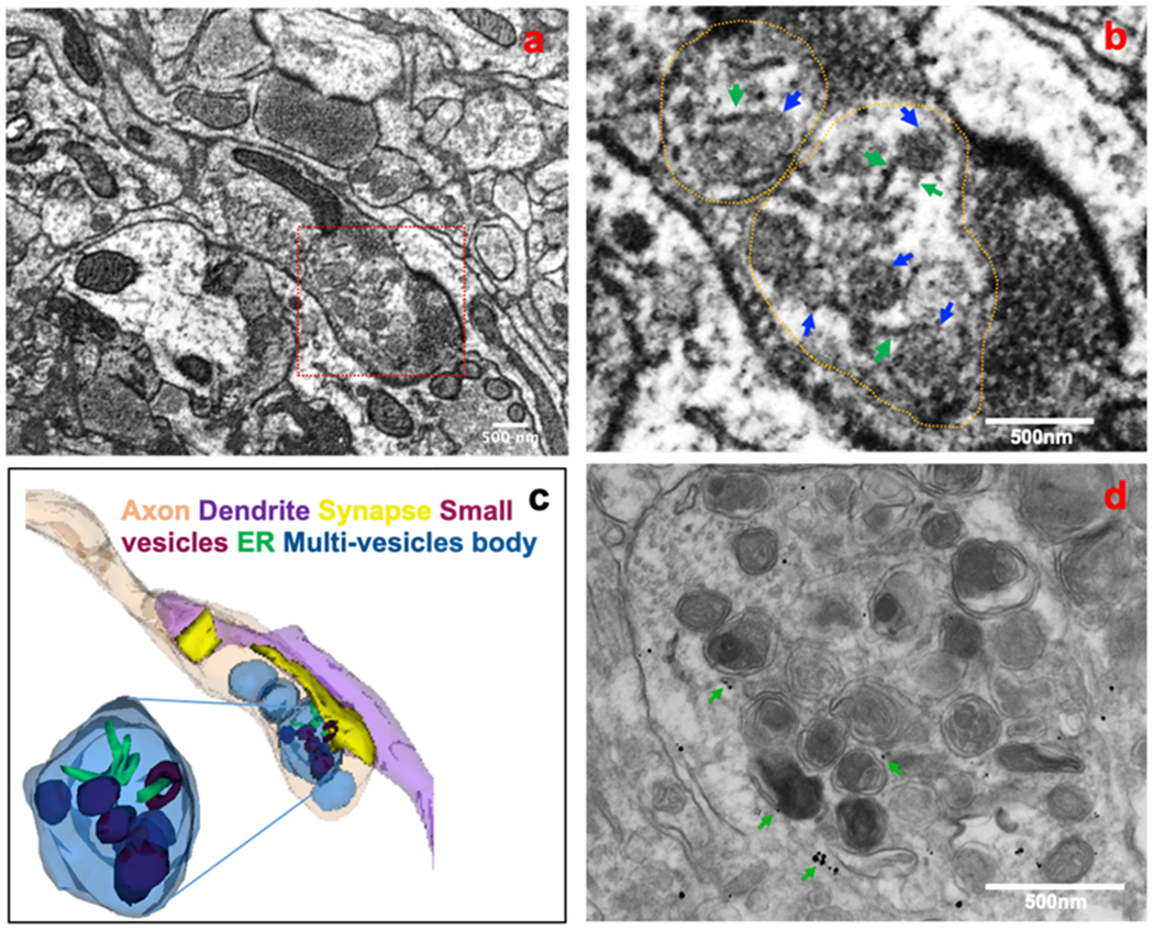
A fixed brain sample from a 5xFAD mouse was subjected to 3D electron microscopy (3D EM). An electron micrograph of a 3D EM image stack shows a link between ER tubules and vesicles during the formation of multi-vesicle bodies in an axonal terminal (a). Association of ER tubules (green arrows) with vesicles (green arrows) in forming a multi-vesicle body is shown in the enlarged view (b). A 3D structure of tubular and vesicular association in a multi-vesicle body was reconstructed from the 3D EM images (c). The reconstructed model shows that ER tubules and vesicles are localized in the same multi-vesicle body and that they associated with each other at some point. Some vesicles (actual brown color) appeared as blue or deep blue in multi-vesicle bodies due to overlay. Immunogold labeling of a 6-month-old 5xFAD mouse brain showing RTN3-labeled ER tubules (indicated by green arrows) in a multi-vesicle body predominantly filled with autophagy vesicles (d). Many of those ER tubules were associated with the vesicles.