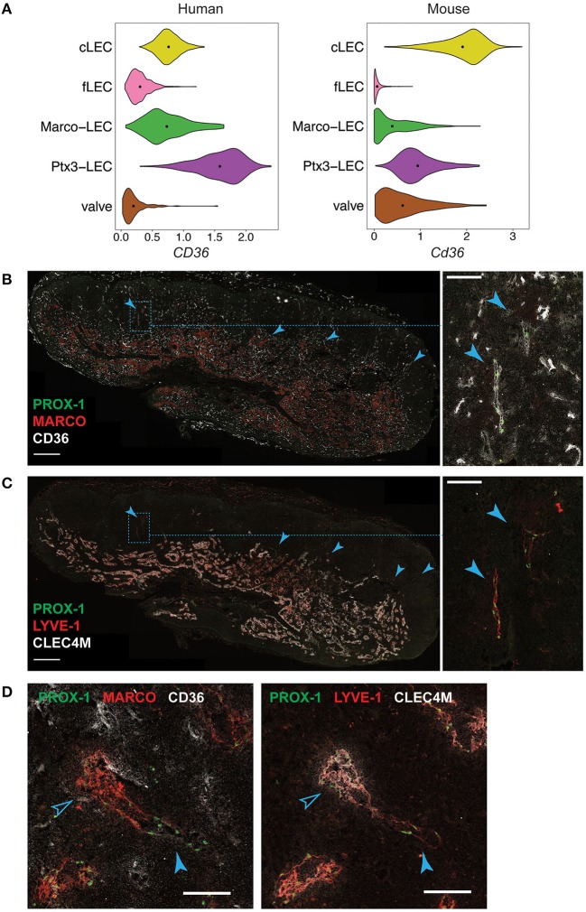
Figure 9.
In situ localization of Ptx3-LECs and transition between Ptx3-LECs and Marco-LECs in human LNs. (A) Expression of CD36/Cd36 in LN LEC subsets of human and mouse. Dots indicate mean log-normalized transcript count. (B–D) Identification of CD36high Ptx3-LECs in human head and neck LNs by immunostaining. (B,C) Immunofluorescence of PROX-1, MARCO and CD36 (B), or PROX-1, LYVE-1 and CLEC4M (C). Zoomed-in images (inset marked by blue dotted lines) in (B) and (C) demonstrate CD36high LYVE-1+ paracortical sinuses (filled arrowhead). Scale bars = 500 μm (left panels) and 100 μm (right panel inset). (D) CD36high LYVE-1+ Ptx3-LECs (filled arrowhead) can be seen associated with MARCO+ CLEC4M+ Marco-LECs (empty arrowhead) in human LNs. Scale bars = 100 μm. CD36high Ptx3-LECs were detected in four out of seven human LNs. Images are representative of four biological replicates.

