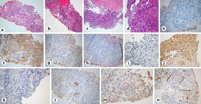
Fig. 2.
a, b H&E stain under ×20 magnification demonstrating a poorly differentiated malignant neoplasm with focal neuroendocrine differentiation in the lung biopsy. c, d H&E stain under ×20 magnification demonstrating a poorly differentiated neoplasm in the duodenal mass biopsy. e–h IHC stains (×20) from the lung. e Negative CK-20 stain. f Negative P-63 stain. g Negative TTF-1 stain. h Negative chromogranin stain. i–l IHC stains from the duodenum (×20). Identical to the lung samples, i shows a negative CK-20 stain, and j shows a negative TTF-1 stain. k Negative CDX-2 stain of the duodenal biopsy. Weakly positive lung biopsy stains (×20) demonstrating CK-7 (l), synaptophysin (m), and calretinin (n). IHC staining of both biopsies were similar; however, ultimately the diagnosis was of undifferentiated histogenesis.

