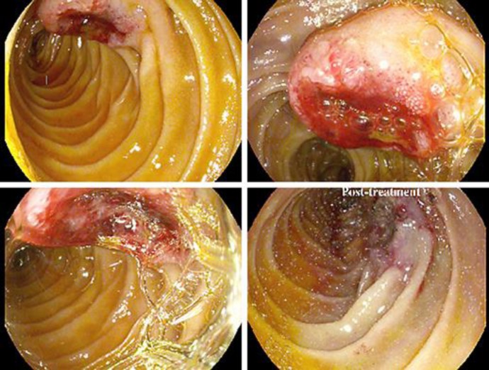
Fig. 3.
Endoscopic images done during the patient's hospital course. The images demonstrate a large, oozing, edematous, heaped-up lesion with an ulcerated center in the third portion of the duodenum. The bottom right image was taken after an endoscopic bipolar electrocautery procedure. Biopsies from the lesion demonstrated metastatic disease histologically similar to his primary lung cancer.

