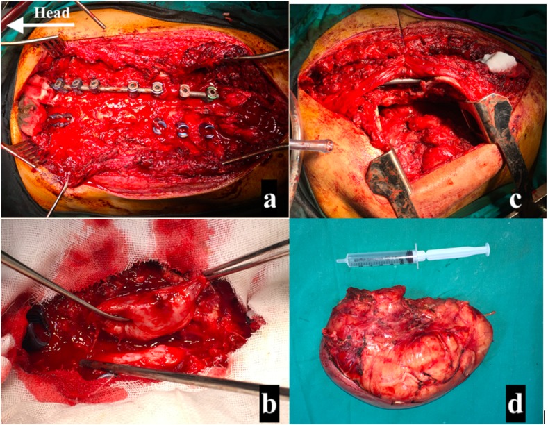
Figure 4.
Intraoperative photos. (a) Pedicle screws insertion with rod connected to the right side and microscopic decompression of the spinal cord. (b) Separation of the mass from the cord and ligation of the connecting nerve root and excision of the mass from the spinal cord. (c) Retraction of the para-spinal muscles medially and entering the left hemithorax through the 6th intercostal space posteriorly. (d) The white tumor mass after complete resection.

