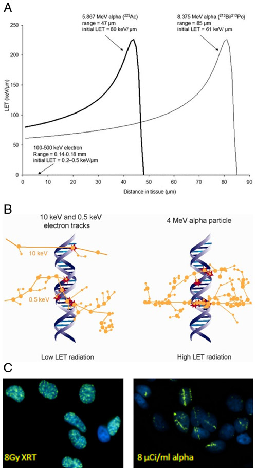
Fig. 3.
Biophysical properties of radiation with different linear energy transfers (LET). A. α-particle LET as a function of distance for α emissions from Actinium-225. The plot shows the LET vs distance track for the 5.9 MeV α-particle emitted directly from 225Ac and also the 8.4 MeV α-particle emitted by bismuth-213, the 45.6 min half-life daughter of 225Ac; a plot of the 0.2 to 0.4 keV/μm LET of electrons is barely visible on the scale of this plot. B. Ionization events (circles) within the DNA molecule (red circles) are illustrated for low LET radiation (10 and 0.5 keV electrons) and for high LET radiation (4 MeV α-particle); figure taken from Ref. [6]. Note that the LET of high energy electrons is lower and would have even fewer DNA interactions than depicted in panel B. C. γH2AX staining in the nuclei of MCF7 cells showing the fluorescence associated with localization of the DNA DSB repair machinery for: left-20 min after 8 Gy (137Cs-irradiator) photon irradiation and right-20 min after incubation with 8 μCi/ml 213Bi-labeled antibody. (from Hong Song, unpublished)

