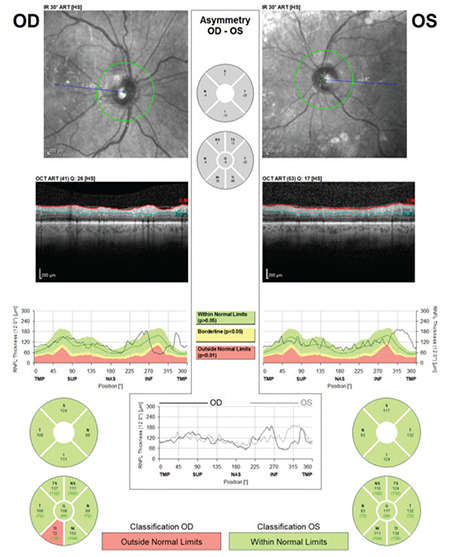
Figure 1.

(Spectralis OCT) Example of a scan with low quality score for the left eye of a patient with cataract. Note that the quality coefficient (Q) is 26 on the right and 17 on the left. In the RNFL profile image of the left eye, RNFL thickness measurements are artificially high in the inferior and temporal regions due to incorrect detection of the RNFL border by the device algorithm
RNFL: Retinal nerve fiber layer, OCT: Optical coherence tomography
