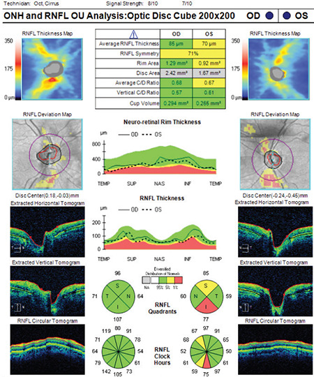
Figure 2.

(Cirrus HD-OCT) Although signal strength is within normal range (≥ 6), there are significant motion artifacts in the deviation map of the right eye (note the breaks in the blood vessels). Similar motion artifacts are also present in the RNFL deviation map of the left eye. Average RNFL thickness is 85 μm in the right eye and 70 μm in the left eye. While the TSNIT profile and RNFL classification are within normal limits in the right eye, there are abnormalities in some sectors of the left eye
RNFL: Retinal nerve fiber layer, OCT: Optical coherence tomography
