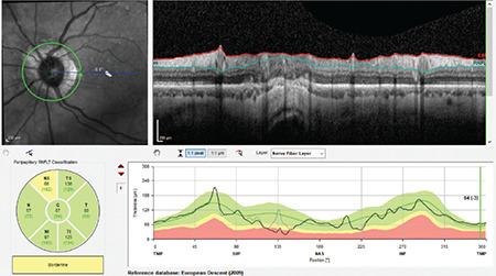
Figure 4.

(Spectralis OCT) Segmentation error resulting from the passage of the scanning ring over an atrophic area in the superonasal region of a myopic patient with peripapillary atrophy. Values on the TSNIT profile are close to zero and peripapillary RNFL classification is borderline in that area
RNFL: Retinal nerve fiber layer, OCT: Optical coherence tomography
