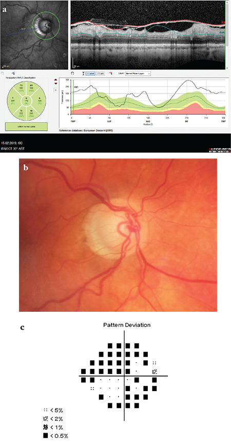
Figure 9.

(Spectralis OCT) Segmentation is completely disrupted in a patient with substantial ILM thickening. Peripapillary RNFL classification appears to be within normal limits in all sectors, and analysis of the sectors shows that average RNFL thickness values are much higher than expected average values. The same findings are seen in the TSNIT profile (a). Disc photography shows prominent glaucomatous pitting and peripapillary atrophy (b). There is significant glaucomatous visual field loss in the same eye (c). Note that OCT RNFL classification appears to be within normal limits in all sectors
RNFL: Retinal nerve fiber layer, OCT: Optical coherence tomography
