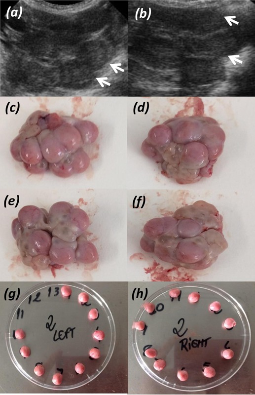
Figure 1.

Example of ultrasound image of the ovaries (a and b, left and right ovary respectively), with the white arrows indicating individual corpus luteum. Panels c and d show the left ovary, and e and f show the right ovary, before dissection. Panels g and h show the individual corpus luteum dissected from left and right ovary, respectively.
