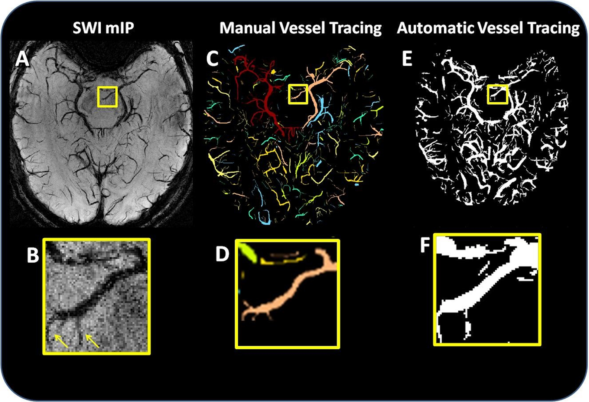
Figure 2:

Comparison of manual and automated segmentation. (A) Axial slice of the SWI mIP, and (B) a magnification of the mIP vessels highlighted in the yellow box. (C) Manually segmented vessels and (D) a magnification of the manual segmentation highlighted in the yellow box. (E) The result of automated vessel tracing, and (F) a magnification of the automatic segmentation highlighted in the yellow box. The arrows in (B) indicate that the automatic segmentation produced accurate segmentations of some mFP vessels (i.e., vessels which were identified in the automated segmentation but not in the manual segmentation).
