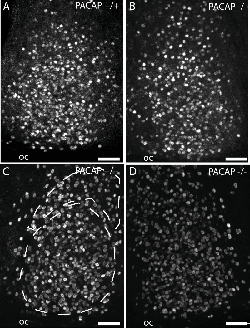
Fig 4. Immunohistochemistry of coronal sections of the mid SCN stained for EGR1 protein at early night (ZT 17).
The core and ventral shell (retinorecipient) and shell portion of the mid SCN [41] is indicated in panel C. Animals were fixed 60 min after a 30 min light stimulation at 300 lux (A and B) or 10 lux (C and D). OC, optic chiasm, Scale bars = 50 μm.

