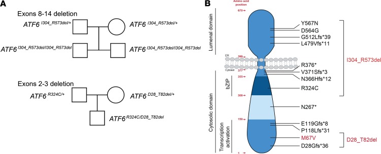
Figure 1. Pedigrees and topography of disease-causing mutations identified in the patients.
(A) Pedigree drawings of patients with deletions of exons 8–14 and exons 2–3 in ATF6. (B) The amino acid organization, protein topology, and functional domains of ATF6 are shown, and the positions of the ATF6 variants are mapped onto the schematic drawing. Variants tested in this study are shown in red.

