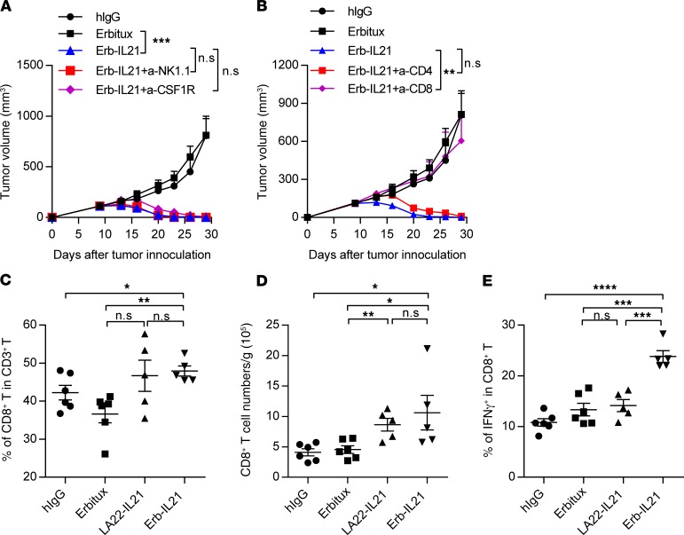
Figure 3. Tumor regression induced by IL-21 depends on CD8+ T cells.
C57BL/6 mice (n = 4–5) were inoculated with 2.5 × 10⁵ MC38-cEGFR cells and were i.p. treated with 75 μg hIgG, Erbitux, or Erb-IL21 on days 10, 13, and 16. (A) Mice were i.p. treated with 400 μg anti-NK1.1 antibody on days 9 and 14 or 300 μg anti-CSF1R on days 9, 12, 15 and 18. One of two representative experiments is shown. (B) Mice were i.p. treated with 200 μg anti-CD4 antibody or anti-CD8 antibody on days 9, 12, 15, and 18. One of two representative experiments is shown. (C–E) Tumor tissues were analyzed 5 days after i.p. treatment of 40 μg hIgG, Erbitux, LA22-IL21, or Erb-IL21 or control antibody. Six hours before sacrifice, mice were i.v. treated with 250 μg BFA to enhance intracellular cytokine staining signals by blocking transport processes during cell activation. (C) Percentage of CD8+ T cells in CD3+ T cells. (D) Total cell number of CD8+ T cells per gram tumor. (E) Percentage of IFN-γ+ in CD8+ T cells. The mean ± SEM values are shown. Two-way ANOVA tests were used to analyze the tumor growth data and unpaired t tests were used to analyze the other data. *P < 0.05, **P < 0.01, ***P < 0.0001, ****P < 0.0001.

