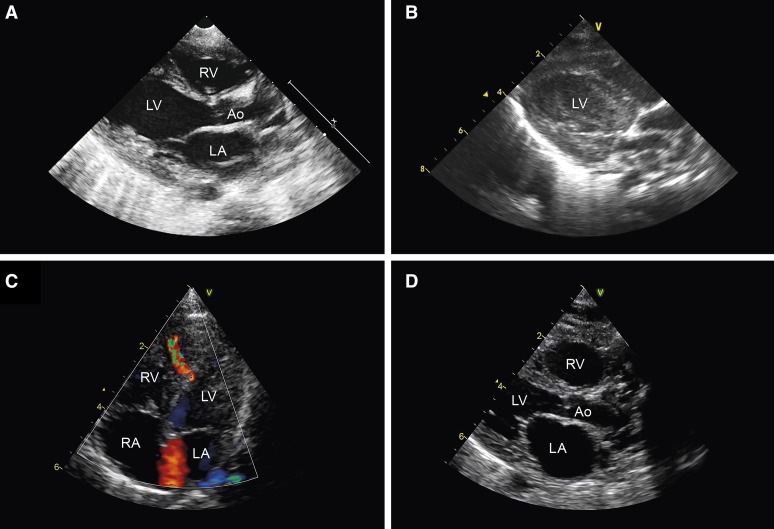
Figure 2.
Cardiac ultrasound examination. Patient II:2 (A) parasternal long-axis view during extracorporeal membrane oxygenation (ECMO) showing mild dilatation of the left ventricle; (B) intracardiac thrombus formation. Note: images from the echocardiogram made before ECMO were not available. Patient II:3 (C) 4-chamber view at first day postpartum showing a midmuscular ventricular septal defect. D, Parasternal long-axis view during cardiopulmonary resuscitation showing dilatation of the heart chambers. Ao indicates aorta; LA, left atrium; LV, left ventricle; RA, right atrium; and RV, right ventricle.

