Figure 3.
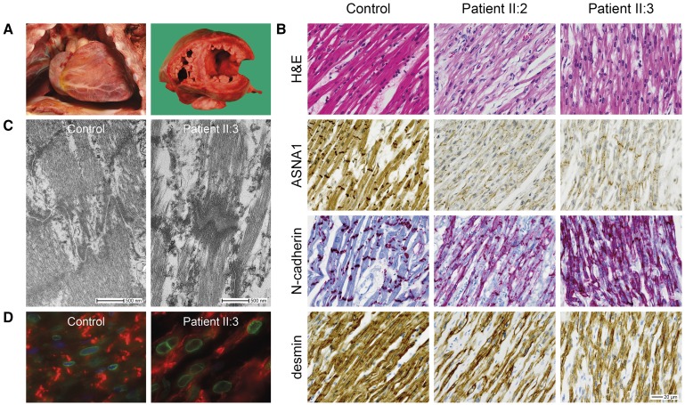
Histopathologic features of the myocardium. A, Macroscopic images showing an enlarged heart with dilated left ventricle in patient II:3. B, Representative images of histological and immunohistochemical studies of myocardial tissue showing markedly reduced expression of ASNA1 at the cytoplasm and intercalated discs in both patients compared with an age-matched control. N-cadherin staining showing irregular appearance of the intercalated discs. Desmin staining showing myofibrillar disorganization. Scale bar: 20 µm. C, Electron microscope images of cardiac intercalated discs, showing increased intercellular space in the patient compared with an age-matched control. D, Representative images of immunofluorescence double-staining of emerin (nuclear membrane, green fluorescence) and N-cadherin (intercalated disc, red fluorescence) in myocardial tissue of patient and control. Note irregular nuclear shape in the patient. Note: experiments were performed in both patients. However, as the images of patient II:2 were of too low quality for publication, only images from patient II:3 are displayed here.

