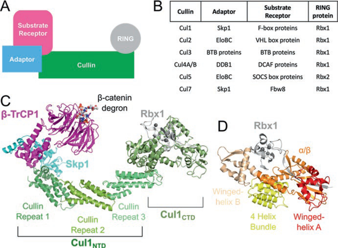
Fig. 12.1.
Architecture of cullin-RING ligases. (a) Schematic representation of the components of a CRL. (b) Specific protein factors for each CRL family. (c) Structural architecture of a CRL1 in ribbon representation generated from PDB IDs 1LDK and 1P22. (d) Organization of Cul1 CTD–Rbx1. The domains of Cul1 are labeled in (c) and (d). The Zn2+ ions in (c) and (d) are shown as gray spheres

