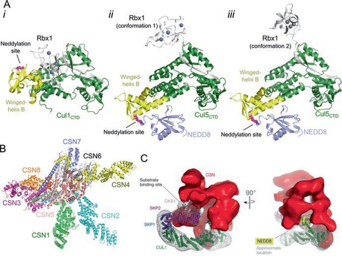
Fig. 12.3.
Neddylation activates cullin-RING ligases. (a) i, Structure of unneddylated, inactive Cul1CTD–Rbx1 (PDB ID: 1LDK). Lys720, the neddylation site, is shown in magenta as sticks. ii and iii, Neddylation of CulCTD causes a change in orientation of the winged-helix B motif and rearrangement of Rbx1 (PDB ID: 4P5O). Shown are two conformations of Rbx1 observed in the crystal structure. The isopeptide bond between Cul5 and NEDD8 is shown as sticks. (b) Crystal structure of the CSN (PDB ID: 4D10). (c) Negative staining electron microscopy reconstruction of CSN bound to CRL1Skp2 (EMD-2173) (Reprinted from Lydeard et al. 2013)

