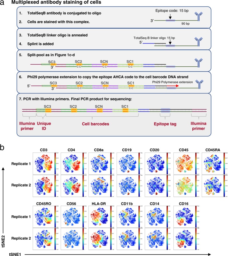
Fig. 5. Protein analysis in human cells.
a Schematic of anti-human antibody QBC workflow. (1–2) TotalSeq-B antibody-oligonucleotide conjugates are stained on cells. (3) A TotalSeq-B linker oligonucteotide is annealed to the oligonucleotide conjugated to the antibodies which contains the “anchor” sequence for later annealing of the Splint. (4–5) Split-pool is done as in Fig. 1. (6) A Phi29 polymerase extension copies the epitope code to the cell barcode DNA strand. (7) PCR with Illumina primers is done to prepare the library for sequencing. b viSNE plots for data from the biological replicates of a 13 anti-human antibody panel applied to human PBMCs. viSNE clustering using 12 out of the 13 markers. CD45 was left out of the clustering analysis to observe how the other leukocyte markers correlated with each other.

