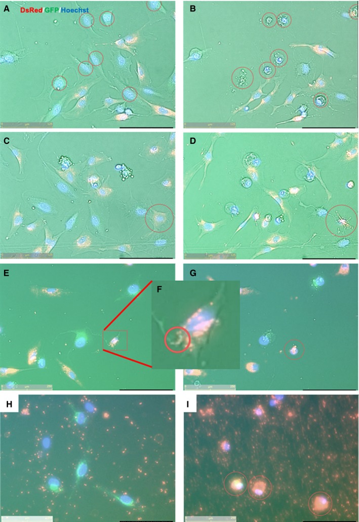
Figure 4.

Continuous fluorescence images of mitochondrial transfers in various conditions. (Recipient cells, EMCs; Mitochondrial donor cells, MSCs). (A, C, E and H) Pre‐culture and (B, D, G and I) post‐culture images. (A&B) Damaged EMC cells exhibiting necroptosis‐like cellular bursts at 24 h following 3 h of H2O2 exposure. (C&D) MSCs which made contact with damaged EMC cells via TNT in the presence of DAMPs were similar in appearance to EMCs as shown in panels A and B, and exhibited necroptosis‐like cellular burst. (E‐G), (F) Enlarged image of E Mitochondrial transfer from damaged EMCs to healthy MSCs via TNT were detected in the presence of DAMPs; consequently, MSCs underwent necroptosis‐like appearance. (H&I) Isolated mitochondria accumulated in damaged EMCs, leading to cell death in the presence of DAMPs. The yellow bar in lower‐left corner indicates 125 µm
