Figure 7.

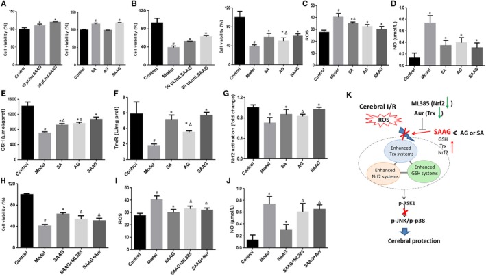
SAAG, SA and AG protected against H2O2‐induced neurotoxicity in PC12 cells; A, Viability of PC12 cells treated with SAAG, SA and AG (n = 5); B, Viability of PC12 pre‐treated with drugs and then followed with H2O2 treatment (n = 6); C, ROS level (n = 6); D, Nitric Oxide level (n = 4); E, GSH level (n = 4); F, TrxR activity (n = 3); G, Nuclear Nrf2 activation (n = 4); H, Cell viability by SAAG was abolished by Aur and ML385; (n = 6), I, Cellular ROS level enhanced by Aur and ML385 (n = 6); J, NO content increased by Aur and ML385 (n = 4); To reveal the role of SA and AG in SAAG, 0.6 mg/mL AG and 10 mg/mL SA was compared with 20 μL/mL SAAG in H2O2‐induced cell injury model, since every millilitre of SAAG contains 30 mg AG and 0.5 g SA according to its instructions; K, Schematic diagram illustrating that SAAG simultaneous enhanced the Trx, GSH and Nrf2 systems against cerebral I/R injury through ASK1‐JNK/p38 cascades. # P < .05 compared with the control group, *P < .05 compared with the model group, and ∆ P < .05 compared with the SAAG group
