Fig. 6.
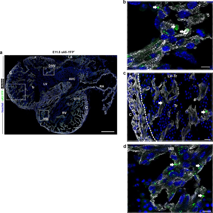
Localization of PCD cells in the E11.5 mouse heart. a Frontal section of a transgenic heart at E11.5. Labeled sA5-YFP PCD cells and bodies (white arrows) were found at the trabeculae of both ventricles, in the regions of the developing mitral and tricuspid valves, in the intraventricular septum, and in the moderator band. b–d Magnifications of the boxed areas in a depicting the presence of PCD (sA5-YFP, arrows). Note the striation of cardiomyocytes due to α-actinin staining. Hoechst nuclear signal, sA5-YFP secreted human Annexin V-yellow fluorescent protein, αActinin cardiomyocyte marker, V ventricle, AVC atrioventricular cushion, RA right atrium, LA left atrium, Tr trabeculae, C compact myocardium, MB moderator band (connects the iVS to the anterior papillary muscle), DTV developing tricuspid valve, and DMV developing mitral valve. Bars are 200 µm (a), 10 µm (b, d), and 20 µm (c)

