Fig. 7.
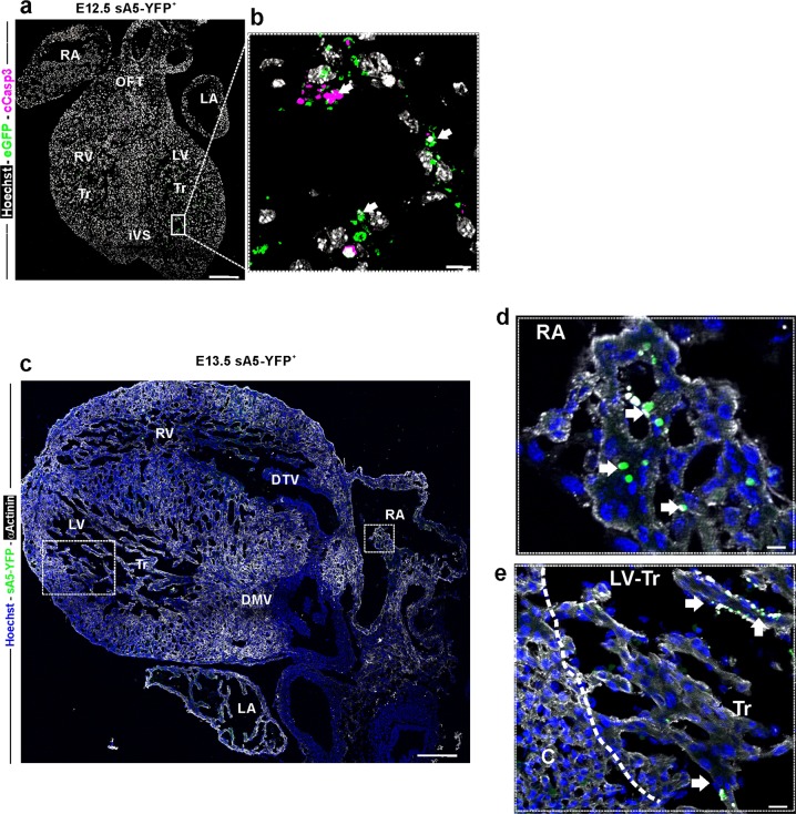
Localization of PCD cells in E12.5 and 13.5 mouse hearts. a, b Sagittal sections of a transgenic heart at E12.5. Magnification of the boxed area in a: bright YFP+ signals (white arrows) mark PCD cells or PCD bodies b. c Frontal sections of a transgenic heart at E13.5. Labeled sA5-YFP PCD cells and PCD bodies (white arrows) were found in the trabeculae of both ventricles, the developing mitral valve, and the right atria. d, e Magnifications of boxed areas in a depicting the presence of PCD (sA5-YFP, arrows). V ventricle, RA right atrium, LA left atrium, Tr trabeculae, DTV developing tricuspid valve, and DVM developing mitral valve. Bars are 200 µm (a, c) and 20 µm (b, d, e)

