Fig. 1.
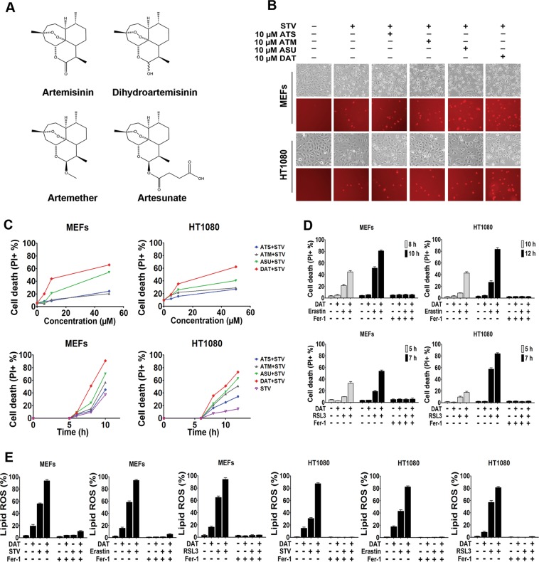
Artemisinin and its derivatives enhance the sensitivity of cells to ferroptosis. a Chemical structure of four artemisinin compounds. b, c In b, ferroptosis was induced in MEF and HT1080 cells by cystine starvation for 8 and 10 h respectively with or without the indicated concentrations of ART compounds. Upper panel shows phase-contrast images and the lower panel shows propidium iodide (PI) staining to indicate dead cells at ×10 magnification. c Flow cytometric quantification of PI positive ferroptotic MEF and HT1080 cells treated with cystine starvation media for 6 and 8 h respectively (upper panel) or with 10 μM of artemisinin compounds (lower panel). d PI positive cells were quantified by flow cytometry after treatment with erastin or RSL3 for the indicated times. e Ferrostatin blocked DAT-sensitized lipid ROS, quantified using BODIPY-C11 lipid probe using flow cytometry in MEF and HT1080 cells. For d and e, cells were treated with 10 μM DAT, 1 μM erastin, 0.5 μM RSL3, and 1 μM Ferrostatin-1. Error bars indicate standard deviation (n = 3)

