Fig. 2.
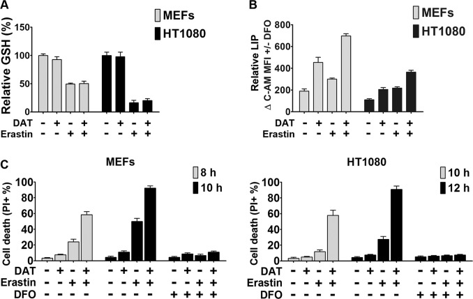
DAT increases cellular free iron contents but has no effect on cellular glutathione. a Quantification of the reduced cellular GSH levels using the DTNB method by measuring optical density at 412 nm after erastin-induced ferroptosis with and without DAT. Values were plotted as a percentage relative to the untreated control sample. b Quantification of cellular LIP levels using the calcein-AM (C-AM) method by measuring spectrophotometric absorbance at 525 nm. The mean fluorescence intensity (MFI) of C-AM is subtracted from the MFI of C-AM treated with DFO. c Cell death was measured by quantifying the percentage PI positive ferroptotic MEF and HT1080 cells by flow cytometry. For a–c, 10 μM of DAT, 1 μM of erastin and 80 μM of DFO concentrations were used for the indicated times

