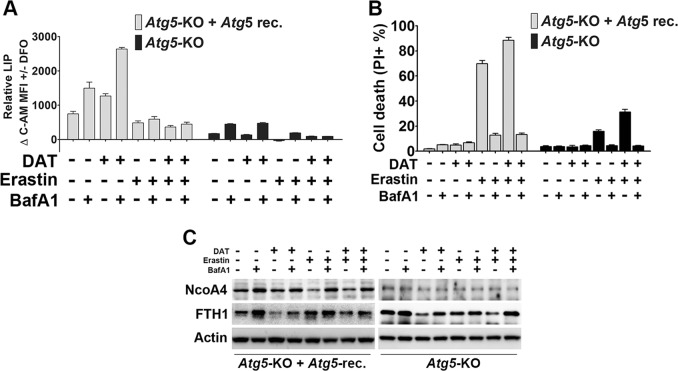
Fig. 4.
Lysosomal activity is required for DAT-sensitized ferroptosis independent of autophagy. a Cellular LIP levels were quantified after treatment with erastin, DAT and/or BafA1 using the calcein-AM method by measuring spectrophotometric absorbance at 525 nm. The MFI of C-AM is subtracted from the MFI of C-AM treated with DFO. b Percentage cell death was quantified by flow cytometric analysis of PI-positive cells in autophagy-deficient Atg5-KO and autophagy-competent Atg5-reconstituted MEF cells after treatment with erastin, DAT and/or BafA1. c DAT can induce the lysosomal degradation of ferritin and increase cellular free iron. Western blot of NcoA4 and ferritin (FTH1) protein expression in Atg5-KO and Atg5-reconstituted MEFs treated with DAT, erastin or BafA1 for 6 h. For a–c, cells were treated with 10 μM DAT, 1 μM erastin or 20 nM BafA1

