Fig. 1.
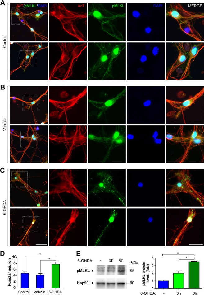
pMLKL activation in cultured neurons after 6-OHDA treatment. a–c Mesencephalic primary cultures were treated with 6-OHDA or vehicle for 1 h. Untreated cultures were used as control. Cells were immunostained against Acetylated Tubulin (AcT, red) and phospho-MLKL (pMLKL, green). Nuclei were stained with DAPI (blue). d Quantification of the number of pMLKL-positive puncta per neuron was estimated in each condition. e Left, cortical primary cultures were treated with 6-OHDA for 3 or 6 h. pMLKL expression was measured by western blot. Hsp90 was used as loading control. e Right, densitometric analysis was performed in each condition for pMLKL and normalized against Hsp90. Scale bar, 30 µm; insets, 15 µm. Data are shown as mean ± SEM. Statistical differences were obtained using one-way ANOVA followed by Bonferroni’s post hoc test. *p < 0.05, **p < 0.01. n = 3 per group

