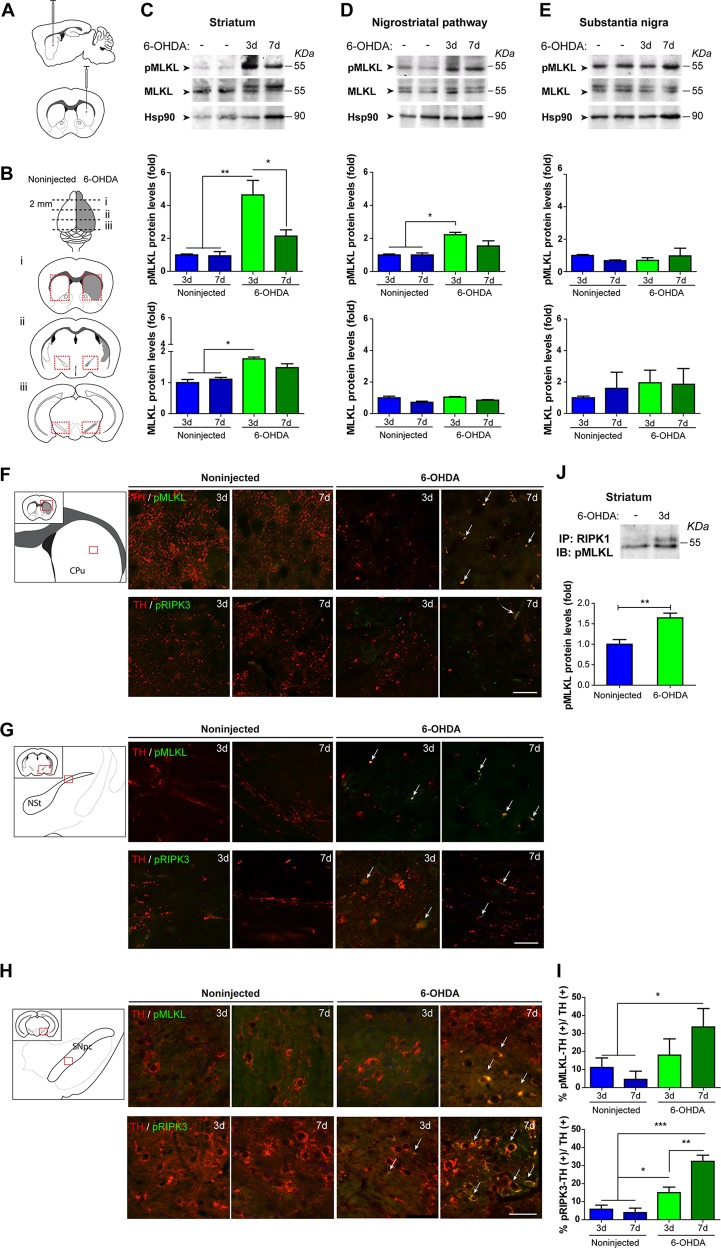
Fig. 4.
Activation of necroptosis in the nigrostriatal pathway after 6-OHDA treatment in vivo. a Wild-type mice were injected with 6-OHDA in the right striatum and analyzed at 3 or 7 days post injection. b Coronal sections of 2 mm thickness were obtained from the i striatum, ii axonal tract, and iii substantia nigra, and the noninjected and injected regions were divided for western blot analysis (indicated in red boxes). pMLKL and MLKL protein expression was evaluated in c striatum, d nigrostriatal pathway, and e substantia nigra. Hsp90 was used as loading control. Coronal sections of f striatum, g nigrostriatal pathway, and h substantia nigra from mice injected with 6-OHDA were immunostained at 3 and 7 days post injection. Schemes show the analyzed area in each brain region. (Upper panels) Sections were immunostained for TH (red) and pMLKL (green) and (Lower panels) TH (red) and pRIPK3 (green). Colocalization in each condition is pointed with arrows. i TH-positive neurons also immunoreactive for pMLKL or pRIPK3 were counted and normalized to the total dopaminergic TH-positive neurons in substantia nigra. j Proteins extracted from 3 days injected striatum and contralateral hemisphere were immunoprecipitated with an antibody against RIPK1 and probed for pMLKL. Relative pMLKL levels were calculated by densitometry. Scale bars, 50 µm. Data are shown as mean ± SEM. Statistical differences were obtained using one-way ANOVA followed by Bonferroni’s post hoc test in c, d, e, and i and by student’s t-test in j. *p < 0.05; **p < 0.01, ***p < 0.001. n = 3 animals per condition

