Fig. 1.
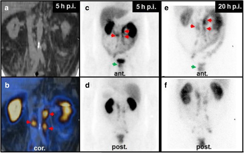
SPECT/CT at 5 h post injection (p.i.) and pairs of ventral/dorsal planar images at 5 h and 20 h p.i. of [99mTc]Tc-PSMA-I&S. a, b SPECT/CT suggested paraaortic lymph node metastases, but the planar scintigraphy (c, d) shows significant hepatobillary and gastrointestinal uptake and suggests urinary secretion. To further distinguish between tumor uptake in paraaortic lymph nodes and unspecific uptake in the ureter (red arrows), a late planar image was taken. e, f Planar scintigraphy at 20 h p.i. Paraaortic uptake remained high and was thus classified as lymph node metastases (red arrows). Note: there was a tracer accumulation in the primary tumor over time, resulting in a better contrast between unspecific bladder signal and the [99mTc]Tc-PSMA-I&S uptake in the prostate (green arrows). Late imaging was restricted to a single bed planar scintigraphy because the low activity at 20 h p.i. would have led to inappropriately high examination times (~ 60 min for planar whole body scintigraphy and whole body SPECT/CT)

