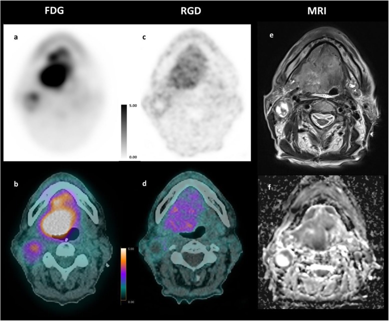
Fig. 4.
Comparative MRI, 18F-FDG, and 68Ga-NODAGA-RGD PET/CT of patient #8. Axial PET/CT fusion slices of a 69-year-old man with a moderate differentiated base tong SCC. The images show different tumor-to-background ratios in between the two radiotracers 18F-FDG PET/CT (a, b) vs. 68Ga-NODAGA-RGD PET/CT (c, d), and also a slightly different distribution of activity within the tumor bed when compared with the MR images e T2w and f ADC map of diffusion

