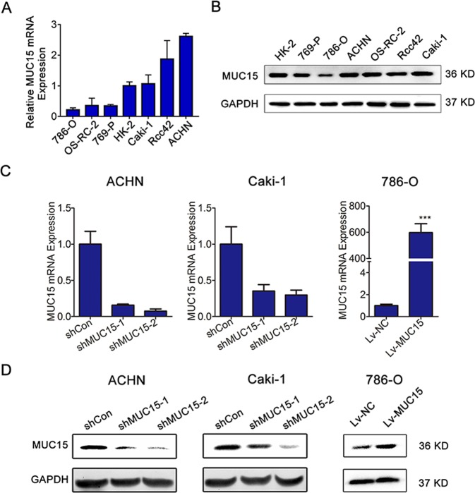
Fig. 2. Expression of MUC15 in RCC cell lines and establishment of sublines.
a, b Quantitative real-time RT-PCR and western blot analysis of MUC15 expression level in human normal and renal cancer cell lines (N = 3). c, d Quantitative real-time RT-PCR and western blotting analysis of MUC15 mRNA expression in ACHN or Caki-1 cell lines transfected with MUC15 shRNAs and shControl, and 786-O cell line infected with MUC15 lentivirus and negative control. 18S was applied as the endogenous control for quantitative real-time RT-PCR, and GAPDH was used as a loading control for western blotting assay (N = 3).

