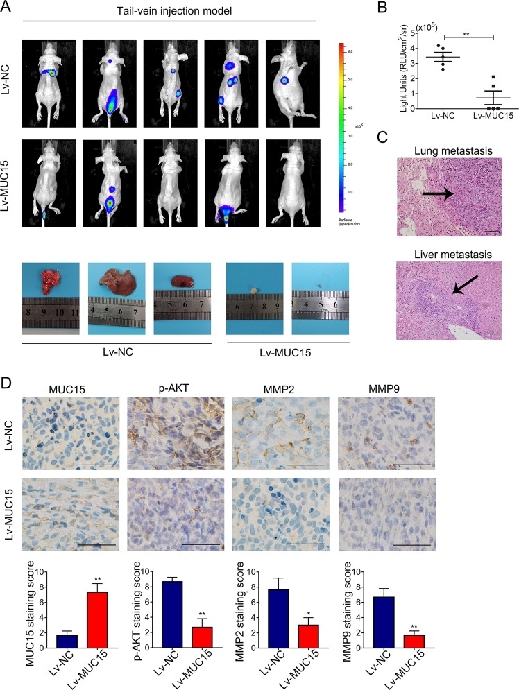
Fig. 6. MUC15 modulated RCC cells distant metastasis in vivo.
a BLI images of athymic BALB/c nude mice implanted with 786-O/NC and 7860/MUC15 cells by tail-vein injection, the images of lung, liver and adjacent metastasis were showed below (N = 5). b Quantification of light emission for metastases in mice (N = 5). (**P < 0.01). c Representative images of Hematoxylin- eosin (HE) staining of lung and liver metastatic tumor (N = 3). d Representative images of immunohistochemistry staining of MUC15, p-AKT, MMP2 and MMP9 in distant or adjacent metastatic tissues from 786-O/NC and 7860/MUC15 cells (N = 3). The scale bar represents 50 μm.

