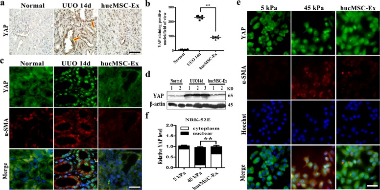
Fig. 3. HucMSC-Ex attenuated tubulointerstitial fibrosis by inhibited YAP.
a YAP immunohistochemistry staining of 14d UUO kidney with hucMSC-Ex treatment. Bar=100 μm. b Quantification of YAP-positive nuclei per field of view from the a experiment. c Double immunofluorescent staining of YAP and α-SMA in the kidney of 14d UUO rats treated with hucMSC-Ex (Green: YAP; red: α-SMA). Bar = 50 μm. d Western blot analyses of YAP expression in the kidney of 14d UUO rats with hucMSC-Ex intervene (n = 3). e The co-localization of YAP and α-SMA in NRK-52E cells cultured under stiff gel condition in the presence of hucMSC-Ex was detected by double immunofluorescent staining. Bar=25 μm. f Statistical analysis the expression of YAP in cytoplasm and nuclear with hucMSC-Ex therapy. **P < 0.01.

