Fig. 1.
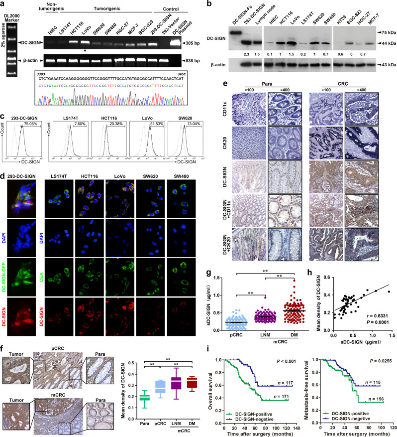
DC-SIGN is frequently upregulated and its positive expression is associated with poor prognosis in CRC. a Transcripts expression of DC-SIGN in various human cancer and normal cell lines was detected by standard PCR (upper panel). The PCR products from LoVo cells were confirmed by sequencing (lower panel). b Expression of DC-SIGN protein in human CRC. HEK293 transfected with DC-SIGN vector, rhDC-SIGN-Fc, and lymph node lysates were used as positive controls. c Cell surface expression of DC-SIGN in colon cancer cell lines was examined by flow cytometry. Numbers listed were percentage of positive cells. Thick line, DC-SIGN; Thin line, isotype control. d Confocal microscopy to determine colocalization of DC-SIGN and CEA in colon cancer cells. e IHC staining of DC-SIGN and CD11c or cytokine 20 coexpression in CRC tissue. f Images shown are representative of DC-SIGN staining in primary (pCRC) and metastatic CRC (mCRC). Para, paracarcinoma; LNM, lymph node metastasis; DM, distant metastasis. g Soluble DC-SIGN (sDC-SIGN) levels in serum derived from CRC patients. h The correlation between the tissue and serum DC-SIGN expression in matched CRC patients. i Kaplan–Meier analysis of the overall survival and metastasis-free survival of CRC patients. Data, mean ± SD. **P < 0.01

