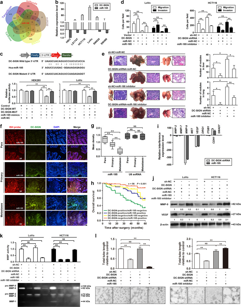
Fig. 3.
miR-185 suppresses MMP-9 and VEGF expression and metastatic potential of CRC cells by targeting DC-SIGN. a Five independent miRNA target databases were used to predict the potential miRNAs. b Endogenous expressions of DC-SIGN and miR-185 in various human CRC cells were detected by qPCR. c Luciferase activity assay for targeting sequences of the 3′-UTR of DC-SIGN by miR-185 in HEK293 and LoVo cells. d Cells were transfected as indicated, and applied to transwell analysis. e The spleens, livers, and lungs were examined for tumors and metastases of the indicated cells (left panel). Quantitation of micrometastases per liver and lung as assessed (right panel). f Representative images of fluorescent in situ hybridization and immunofluorescence of miR-185 and DC-SIGN in paired CRC tissues. Para, paracarcinoma. U6 snRNA was used as a control. g Expression of miR-185 and U6 were semiquantitative by ISH analysis. h Kaplan–Meier curves for all patients divided by combination of miR-185 and DC-SIGN status. i Relative mRNA levels of CRC metastasis-related genes in DC-SIGN-depleted LoVo cells. j The lysates of stable LoVo and HCT116 cells were applied to western blot. k Culture supernatants of CRC cells were applied to gelatin zymography. l Representative images of stable LoVo and HCT116 tube formation of cells on the Matrigel. Data, mean ± SD. *P < 0.05, **P < 0.01. N.S., nonsignificant

