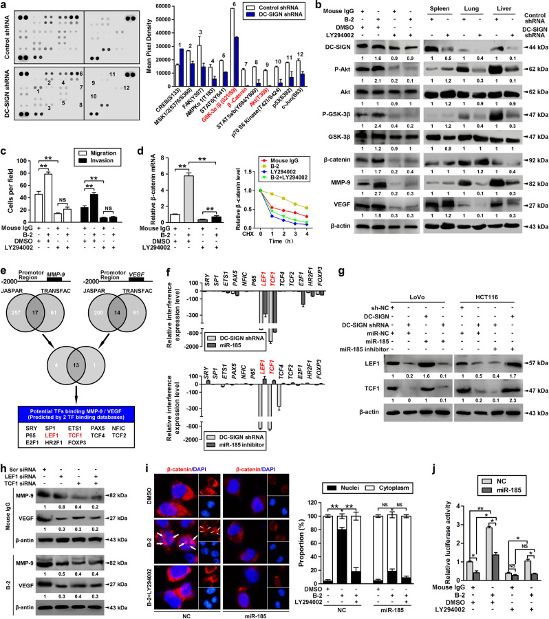
Fig. 4.
miR-185/DC-SIGN signaling blocks β-catenin translocation through the PI3K/Akt/GSK-3β pathway. a The lysates of stable LoVo cells infected with Lenti-DC-SIGN shRNA or control were applied to phosphokinase antibody array, and pixel densities of indicated proteins were shown. b LoVo cells were treated with DC-SIGN agonistic antibody (B-2, 10 μg/ml) for 15min and/or PI3K inhibitor (LY294002, 50 μM) for 24h, followed by western blot analysis (left panel). Stable LoVo cells expressing control shRNA or DC-SIGN shRNA were injected into the spleen. The lysates of spleen, lung, and liver tissues were applied to western blot (right panel). c LoVo cells were treated with DC-SIGN mAb and/or LY294002, and applied in transwell analysis. d Total RNA was extracted and applied to qPCR (left panel), or cells were further pulsed with CHX (20 μM) and applied to western blot (right panel). e Two independent TFs target databases were used to predict the potential TFs binding to promotors of MMP-9 and VEGF. f Interference levels of indicated TFs were shown by qPCR. g Cells were transfected as indicated, and lysates were applied to western blot. h LoVo cells expressing control LEF1 and/or TCF1 siRNA were treated with nonspecific IgG or DC-SIGN mAb. Then cell lysates were applied in western blot analysis. i Immunofluorescent analysis of β-catenin subcellular distribution in LoVo cells transfected with miR-185 or control while being treated with DC-SIGN mAb and/or LY294002 (left panel). Quantitation of β-catenin distribution (right panel). j LoVo cells were treated as indicated, and applied to luciferase-based β-catenin/TCF transcriptional activity assay (TOPflash/FOPflash). Data, mean ± SD. *P < 0.05, **P < 0.01

