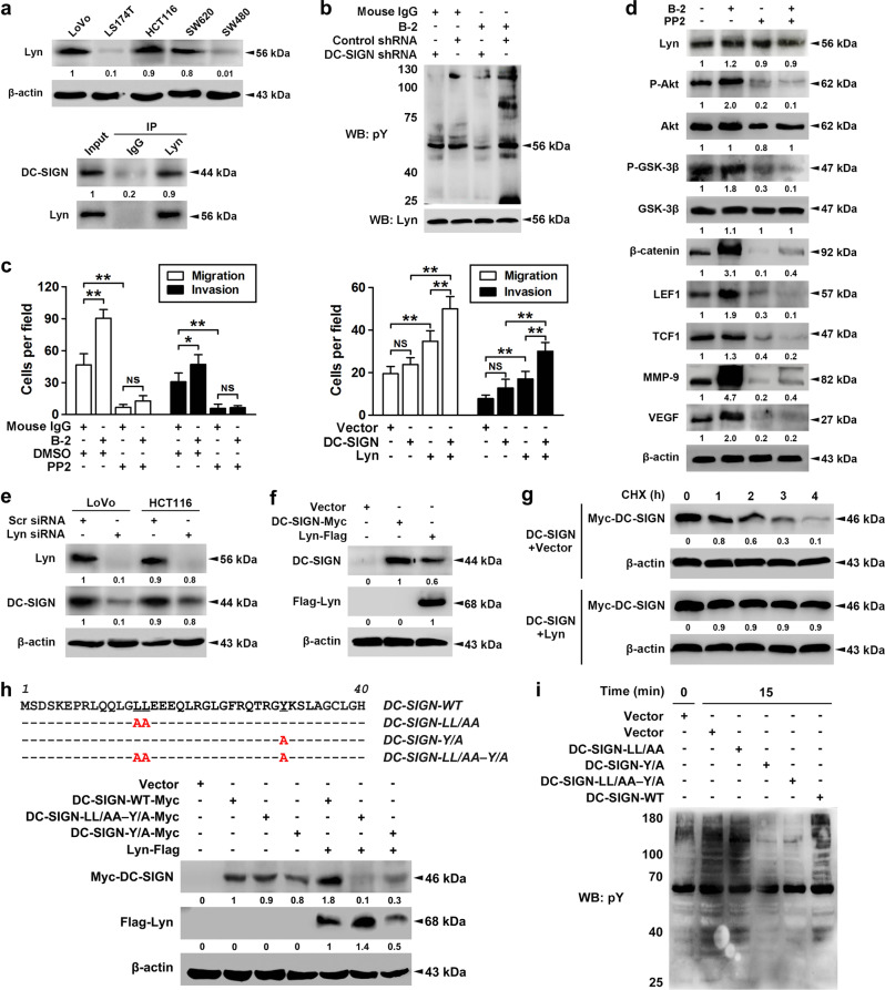
Fig. 5.
DC-SIGN promotes the CRC metastasis through PI3K/Akt/β-catenin activation in tyrosine kinase Lyn-dependent manner. a The expression profiles of Lyn in CRC cells were shown in upper panel, and immunoprecipitation using Lyn antibody was performed in LoVo cell lysates (lower panel). b Stable LoVo cells treated with nonspecific IgG or DC-SIGN mAb (B-2, 10 μg/ml) were collected, lysed, and cell lysates were applied to immunoprecipitation with Lyn antibody. c LoVo cells treated with DC-SIGN mAb and/or PP2 (50 μM, left panel) and LS174T cells infected with DC-SIGN and/or Lyn vectors (right panel), and applied in transwell analysis. d LoVo cells were treated with DC-SIGN mAb and/or PP2. Then cell lysates were applied in western blot analysis. e LoVo and HCT116 cells were transiently transfected scramble or Lyn siRNA, and lysates were applied to western blot. f LS174T cells were transfected with vector, Lyn, or DC-SIGN construct, and lysates were applied to western blot. g LS174T cells were transfected as indicated, pulsed with cycloheximide (CHX, 20 μM), and applied to western blot. h Amino acid sequence alignment of the cytoplasmic domain of WT and mutant DC-SIGN proteins (upper panel). Red letters identify amino acid substitutions. LS174T cells were transfected with constructs as indicated, followed by western blot (lower panel). i After transfection, cells were treated with DC-SIGN mAb, and lysates were applied to western blot. Data, mean ± SD. *P < 0.05, **P < 0.01. N.S., nonsignificant

