Fig. 2.
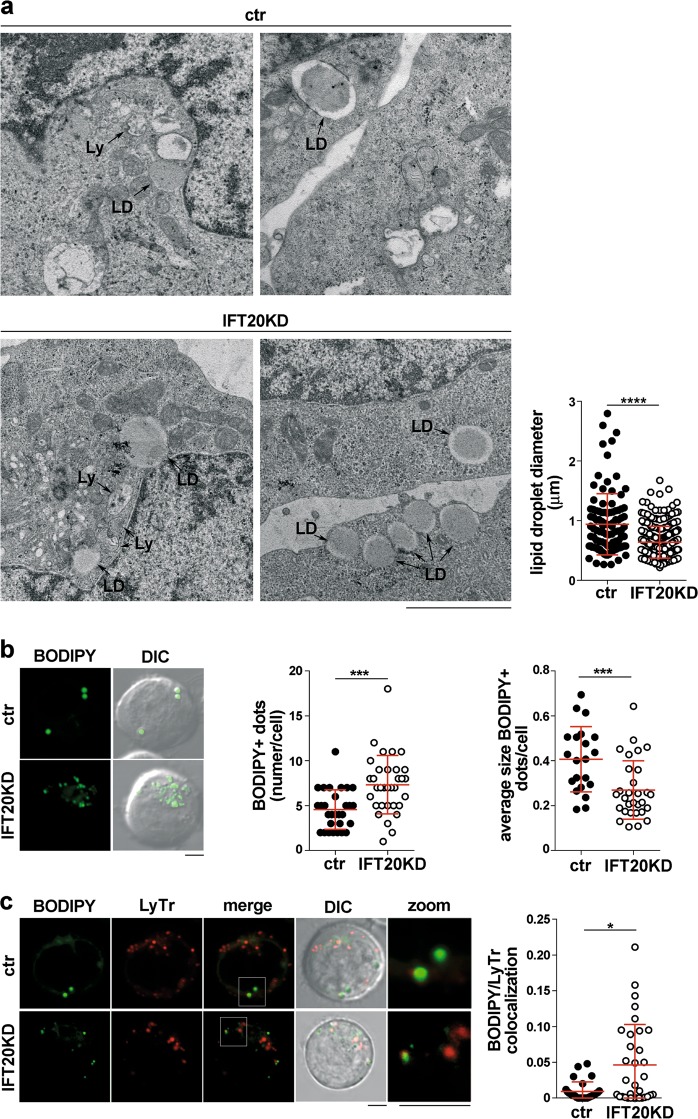
IFT20 is required for lipid droplet breakdown. a Transmission electron microscopy of ctr and IFT20KD Jurkat cells. Lysosomes (Ly) and lipid droplets (LD) are indicated. The graph shows the quantification of the diameter of lipidic droplets in ctr and IFT20KD Jurkat cells (n = 3). Size bar: 2 μm. b Fluorescence analysis of ctr and IFT20KD Jurkat cells loaded with the LD marker BODIPY. Representative images (medial optical sections) are shown on the left. Size bar: 5 μm. The graphs show the quantification of the number of BODIPY+ dots/cell (middle) and the average size of BODIPY+ dots (right) (mean ± SD). At least 21 cells from three independent experiments were analyzed. c Immunofluorescence analysis of ctr and IFT20KD Jurkat cells co-labeled with BODIPY and Lysotracker Red (LyTr). Representative images (medial optical sections) are shown on the left. Size bar: 5 μm. The graph shows the quantification of the colocalization of BODIPY and LyTr, expressed as Mander’s coefficient (mean ± SD). At least 28 cells from three independent experiments were analyzed. *P < 0.05; ***P < 0.001 (Mann–Whitney test)

