Fig. 5.
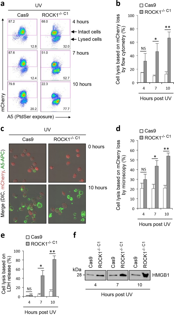
Loss of ROCK1 expression promotes early onset of secondary necrosis. a Flow cytometry analysis of intact (mCherryHigh) and membrane permeabilised (mCherryLow) Cas9 and ROCK1−/− C1 cells 4, 7 and 10 h post UV irradiation. Levels of cell lysis over time were quantified based on the loss of mCherry in cells by flow cytometry (b) and live microscopy (c and d), as well as the release of LDH (e) and HMGB1 (f) into the culture supernatant. b, d and e Error bars represent s.e.m. (n = 3). Data are representative of at least three independent experiments. *P < 0.05, **P < 0.01, NS = P > 0.05, unpaired Student’s two-tailed t-test

