Fig. 3.
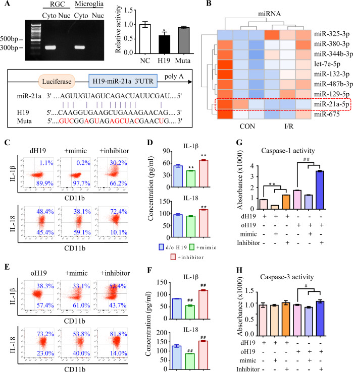
H19 sponges miR-21 to regulate neuro-inflammation. a As measured by qPCR, H19 was specifically expressed in the cytoplasm instead of the nucleus in both RGCs and microglia. Compared to NC RNA, miR-21 reduced luciferase activity by 43.4% ± 1.4% through binding to H19. The miR-21-dependent inhibition of luciferase activity was abolished after mutations (red) in the binding site. Data were represented as means ± SD (n = 3). Compared with NC RNA: *P < 0.05. b The top nine dysregulated miRNAs in I/R retinas. c, d Anti-inflammatory effect of H19 excision was further promoted by the miR-21 mimic with less IL-1β and IL-18 production in supernatant of retinal microglia. As measured by flow cytometry and ELISA, the miR-21 inhibitor greatly antagonized the anti-inflammation effect of dH19 with more production of IL-1β and IL-18. e, f In H19 overexpressed microglia, miR-21 downregulation further aggravated the production of IL-1β and IL-18, which was ameliorated by miR-21 upregulation as measured by flow cytometry and ELISA. g, h Caspase-1 activity was further restrained in the microglia deficient of both H19 and miR-21 compared with the single H19 excision. The miR-21 inhibitor further augmented caspase-1 activity in oH19 microglia. Either H19 or miR-21 had no significant effect on apoptotic activity of caspase-3 in microglia. Data were represented as means ± SD (n = 6). Compared with the dH19 group: **P < 0.01. Compared with the oH19 group: ##P < 0.01. Cyto, cytoplasm; Nuc, nucleus; NC, normal control; Muta, mutation; dH19, H19 knockout; oH19, H19 overexpression

