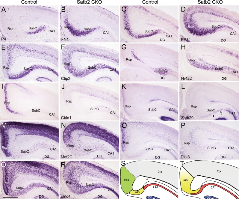
Fig. 4.
Rsp neurons lose their identity in Satb2 CKO mice. FN1 is restricted to the SubC of controls (a) but expressed by neurons in the Rsp region of Satb2 CKO mice at P4 (b). c–h ER81, Citp2, and Nr4a2 are expressed in the SubC and specific layers of the Rsp in control mice (c, e, g). In Satb2 CKO mice, however, they are expressed throughout the Rsp regions while remain largely unchanged in the SubC (d, f, h). Cbln1 shows restricted expression in the Rsp of control mice (i), but its expression is lost in Satb2 CKO mice (j). Zbtb20 is expressed in superficial Rsp and CA1 of control mice (k), but undetectable in the Rsp region while remains unchanged in the CA1 of Satb2 CKO mice (l). Note that ectopic Zbtb20+ cells (arrows) are present in the subicular region of Satb2 CKO mice. Mef2C is intensely expressed in the Rsp and weakly in the SubC of control mice (m), but its expression was largely reduced in the Rsp region to a comparable cellular density of the SubC in Satb2 CKO mice (n). Dkk3 is strongly expressed in upper Rsp of control mice (o) but absent in the Rsp region of Satb2 CKO mice (p). Intense Lmo4 expression is reduced in the Rsp regions with a similar intensity to that in the SubC in Satb2 CKO mice (r) compared with controls (q). s, t Diagram of Rsp fate change in Satb2 CKO mice. DG dentate gyrus. n = 3 mice for each genotype. Scale bar = 500 μm

