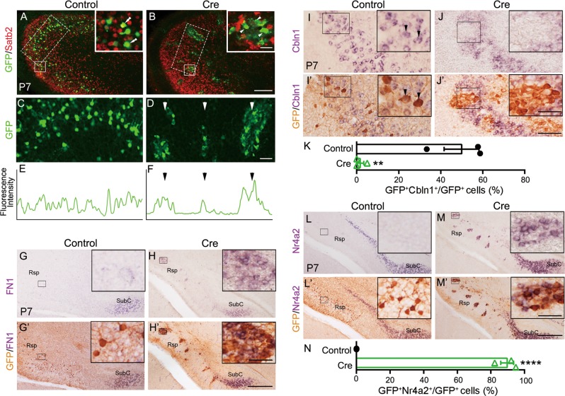
Fig. 5.
Deletion of Satb2 in a subset of Rsp neurons leads to ectopic expression of SubC-specific genes. Rsp neurons of Satb2f/f mice were transfected with GFP alone or GFP plus Cre-expression plasmid by in utero electroporation at E14.5 and analyzed at P7. a GFP-expressing neurons (green) are evenly distributed in the Rsp and adjacent neocortex, and some of them are immunostained with Satb2 antibody (double arrowheads, insert) in Satb2f/f mice. b Cre-expressing (Satb2-mutant) neurons (green) are not immunostained with Satb2 antibody (arrowheads, insert). c–f Knockout of Satb2 in Rsp neurons results in clumping of cell bodies, instead of even distribution. g, h′ Clumping Satb2-mutant neurons initiate expression of the SubC-specific gene FN1 in the Rsp (h′), but control neurons do not (g′). For more clear signals, mRNA signals were photographed first, and then immunostaining of GFP was performed and imaged again. i–j′ Clumping Satb2-mutant neurons located in the middle portion of Rsp fail to express the Rsp-specific gene Cbln1 (j′) but control neurons do so (arrowheads, i′). k Quantitation of Cbln1-expressing GFP+ cells out of total GFP+ cells. About half of GFP+ control cells express Cbln1, while very few GFP+ mutant cells do so. **p < 0.01 (n = 3), error bars represent S.E.M. l–m′ GFP-transfected (control) cells do not have ectopic Nr4a2 expression in the Rsp of Satb2f/f mice (l′), but Cre-expressing Satb2-mutant cells do so m′. n Quantitation of Nr4a2-expressing GFP+ cells out of total GFP+ cells. ****p < 0.0001 (n = 3), error bars represent S.E.M. Scale bars = 50 μm in inserts of b, h′, j′, and m′, 200 μm in b, 50 μm in d, and 400 μm in h′, j′, and m′

