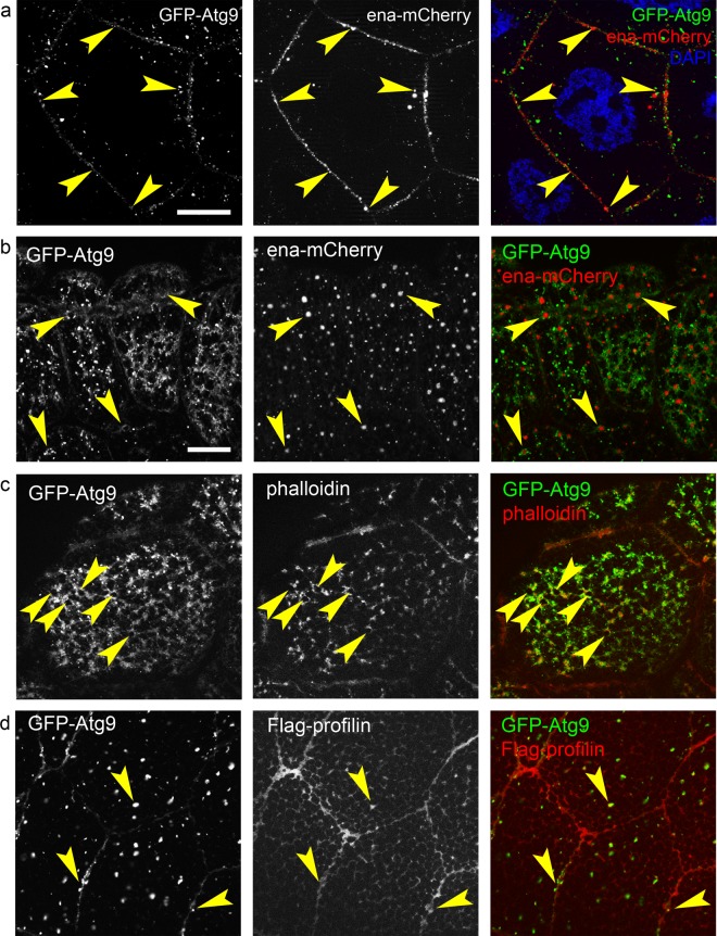
Fig. 6.
Colocalization analysis of Atg9, Ena, and profilin. GFP-Atg9 clearly overlaps with Ena-mCherry along the plasma membrane of nurse cells, and these two proteins are adjacent with partial overlap on punctate cytoplasmic structures (arrowheads in a). GFP-Atg9 (b) and actin phalloidin (c) forms ring-like “nests” (arrowheads) around Ena-mCherry positive structures near the periphery of larval salivary gland cells. GFP-Atg9 overlaps with Flag-profilin along the plasma membrane and on cytoplasmic structures (arrowheads) in larval salivary gland cells (d). Scale bars represent 20 µm in a and b–d

