Fig. 5.
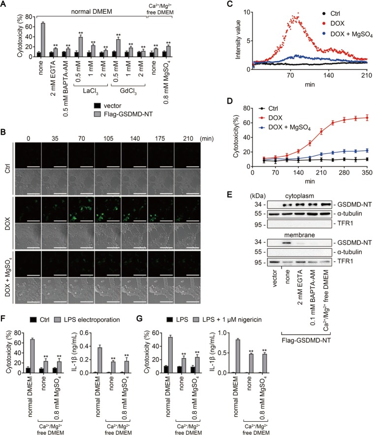
Mg2+ inhibits GSDMD-NT-induced pyroptosis by blocking Ca2+ influx. a, e HEK293T cells transfected with Flag-GSDMD-NT (or empty vector) were treated as indicated. Percentage of LDH release was measured (a), and immunoblots for GSDMD-NT in the cytosolic and membrane fractions were performed (e). Cytosolic and membrane-bound proteins were separated by using a Plasma membrane protein isolation kit. α-tubulin and TFR1 are used as loading controls respectively. b−d HEK293T cells transduced with a doxycycline- (DOX-) inducible human GSDMD-NT cDNA were transfected with GCaMP6 plasmid, a genetically encoded Ca2+ indicator. Cells were treated (or untreated) with DOX 16 h later, in the absence or presence of 10 mM MgSO4. b Representative time-lapse cell images (brightfield and fluorescence) taken from 0 to 210 min after DOX addition. Bar = 100 μm. c Scatter plot of the fluorescence intensity of GCaMP6. d LDH release measured from 35 to 350 min after DOX addition. f iBMDMs primed with Pam3CSK4 overnight were electroporated with (or without) LPS and incubated in the indicated media for 2 h. LDH (left) and IL-1β (right) release were measured. Cells electroporated without LPS (Ctrl) were used as controls. g iBMDMs primed with LPS for 4 h were treated (or untreated) with 1 μM nigericin in the indicated media for 1 h. LDH (left) and IL-1β (right) release were measured. For panels (a, f, and g), data are presented as mean ± SD; **P < 0.01, compared to the cells transfected with Flag-GSDMD-NT (a), electroporated with LPS (f), or treated with nigericin (g) in normal DMEM. Each panel is a representative experiment of at least three replicates. See also Video S1. LPS lipopolysaccharide, iBMDM immortalized bone marrow-derived macrophage, DMEM Dulbecco’s modified Eagle’s medium

