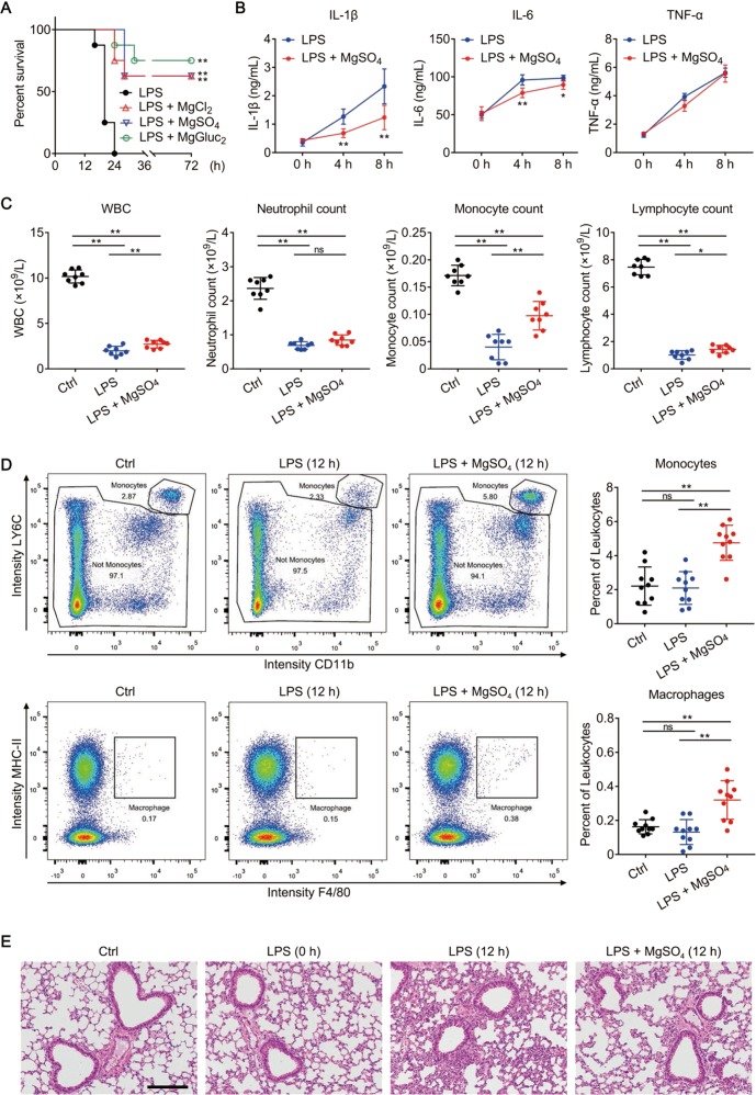
Fig. 7.
Mg2+ is protective in LPS-induced endotoxic shock. a−e C57BL/6 male mice were left untreated (Ctrl group) or primed with 0.4 mg/kg E. coli O111:B4 LPS intraperitoneally and then challenged 6 h later with 10 mg/kg E. coli O111:B4 LPS intraperitoneally. Upon the secondary challenge, LPS was mixed with 1 mmol/kg MgSO4, MgCl2 or MgGluc2 as indicated, or an equal volume of 0.9% NaCl (LPS group). N = 8 per group. a Survival of mice after LPS challenge. **P < 0.01, compared to the LPS group. b IL-1β, IL-6, and TNF-α levels in mouse plasma measured at the indicated time points after LPS challenge. Mice treated with MgSO4 were compared to the LPS group at each time point. c Cell counts of white blood cell (WBC), neutrophil, monocyte, and lymphocyte in mouse whole blood measured in a normal state (Ctrl) or at 12 h after LPS challenge. d Representative flow cytometry plots (left) and quantification (right) of monocytes (CD11b+LY6C+LY6G−) and macrophages (CD11b+MHC-II+F4/80+) in mouse whole blood measured in a normal state (Ctrl) or at 12 h after LPS challenge. e Representative H&E-stained lung sections obtained from mice in a normal state (Ctrl) or at 0 or 12 h after LPS challenge. Bar = 100 μm. Data are presented as mean ± SD. *P < 0.05, **P < 0.01, ns = not significant. Each panel is a representative experiment of at least two replicates. See also Fig. S8

